R&D AF3634, AF3720
For hemojuvelin determination in experimental animals, AF 3634 is the antibody of choice. It detects HJV in whole liver homogenates, liver microsomes and the plasma membrane-enriched fraction of liver homogenate of both mice and rats (Frýdlová J et al., Effect of Erythropoietin, Iron Deficiency and Iron Overload on Liver Matriptase-2 (TMPRSS6) Protein Content in Mice and Rats. PLoS One. 2016 Feb
4;11(2):e0148540). And, although the function of HJV is muscle is dubious, also in muscle (Fujikura Y et al., Liver and muscle hemojuvelin are differently glycosylated. BMC Biochem. 2011 Sep 21;12:52) and the myocardium.
In homogenates and microsomes, the antibody detects mainly a 35 kDa band, which is probably produced by autocleavage at a labile bond in the full-length protein:
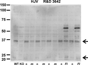
The blot shows on the left liver homogenates (in NP40 buffer) from wild-type (WT) and hemojuvelin knockout mice (KO); the next samples represent NP40 homogenates from control mice (c) and Tmprss6-mutated mice, also known as mask mice (m). The main hemojuvelin bands can be seen at approximately 35 kDa. The two lanes on the right (Fl) represent samples from hemojuvelin-overexpressing mice; in these mice, the hemojuvelin band (including the Flag tag) is seen at approximately 55 kDa.
In the plasma membrane-enriched fraction of mouse liver, HJV is detected as a full-length protein of about 50 kDa. It is correctly glycosylated, as PNGase F treatment shifts the full length protein from 50 kDa to about 35 kDa.
There is a related antibody, AF3720, it seems to give similar results.